APPLICATIONS & THERANOSTICS
J-PET enables and enhances applications across:
- Oncology — staging, recurrence, tumor metabolism
- Neurology — cognitive disorders, Alzheimer’s research
- Cardiology — viability, perfusion, inflammatory disease
- Pediatric Imaging — low-dose protocols
- Targeted Radioligand Therapy (Theranostics)
- Proton Therapy Monitoring
- Dual-Tracer Imaging
Theranostics is rapidly transforming cancer treatment—and J-PET is engineered to unlock imaging capabilities required for expanding alpha- and beta-radiopharmaceutical therapies.
The demonstration of the potential of J-PET technology for theranostics application was demonstrated in the Medical University of Warsaw. Fig. 1 shows the course of diagnosis and treatment of a patient with secondary recurrent glioblastoma with the application of the portable modular J-PET scanner.
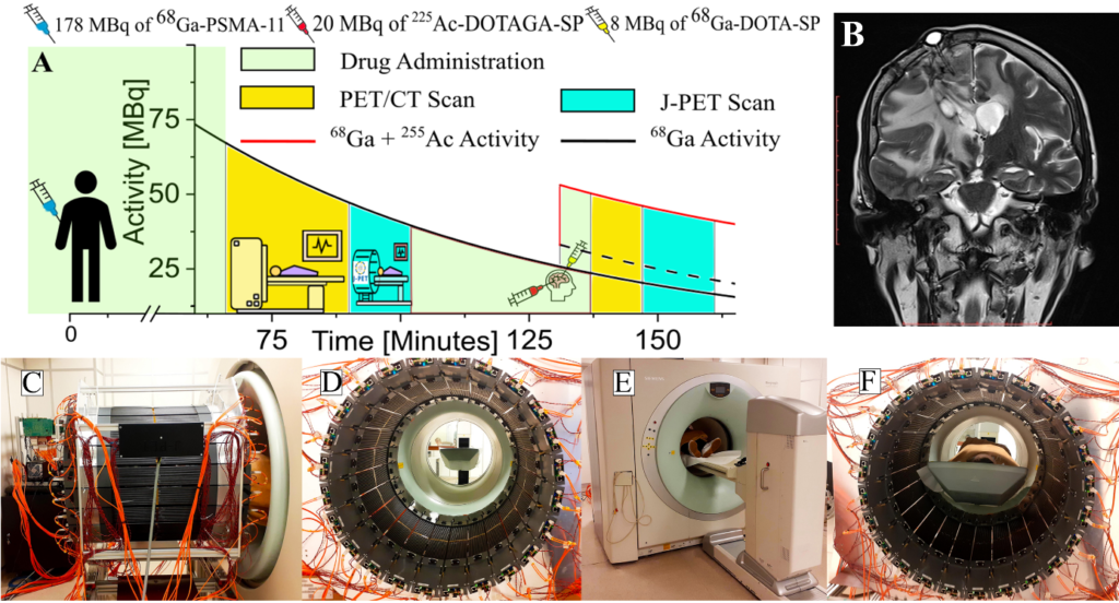
Fig. 1. Course of diagnosis and treatment of a patient with secondary recurrent glioblastoma.
(A) The solid black curve indicates the decrease in the activity of the 68Ga radionuclide after intravenous injection of a 178-MBq activity of the [68Ga]Ga-PSMA-11 radiopharmaceutical followed 131 min later by intratumoral administration of 8 MBq of [68Ga]Ga-DOTA- SP (black dashed curve) together with 20 MBq of [225Ac]Ac-DOTAGA-SP (red curve). After the first and the second administration of pharmaceuticals, the patient was imaged with the Siemens PET/CT Biograph 64 TruePoint and then with a modular J-PET for the time indicated in the graph in yellow and turquoise, respectively.
(B) The T2-weighed MRI coronal image of the head (Magnetom 3T, Siemens Healthcare) showing the tumor and the position of the cat-cath system (visible on the upper left part of the head) for the administration of a radiopharmaceutical for the local treatment of glioblastoma.
(C to F) Photographs illustrating the course of patient imaging with the modular J-PET scanner.
(C) Modular J-PET scanner placed behind the PET/CT Biograph 64.
(D) View of the patient’s bed from the inside of the J-PET tomograph.
(E) The patient was moved on the table so that the torso and legs were in the Biograph PET/CT and the head in the J-PET scanner. In this view, only the patient’s feet are visible on the edge of the Biograph PET/CT. (F) Photograph of the patient with the head inside of the J-PET scanner during imaging after intravenous administration of the [68 Ga]Ga-PSMA-11 radiopharmaceutical.
The figures and caption are taken from the article published in Science Advances where more details about the medical protocol are available.
https://www.science.org/doi/10.1126/sciadv.adp2840
As a result of this study the first ever positronium image was demonsrated as described in Science Adavances published in 2024, and also in the a review on positronium imaging featured in the November 2025 issue of IEEE Transactions on Radiation and Plasma Medical Sciences available via link: IEEE Master email
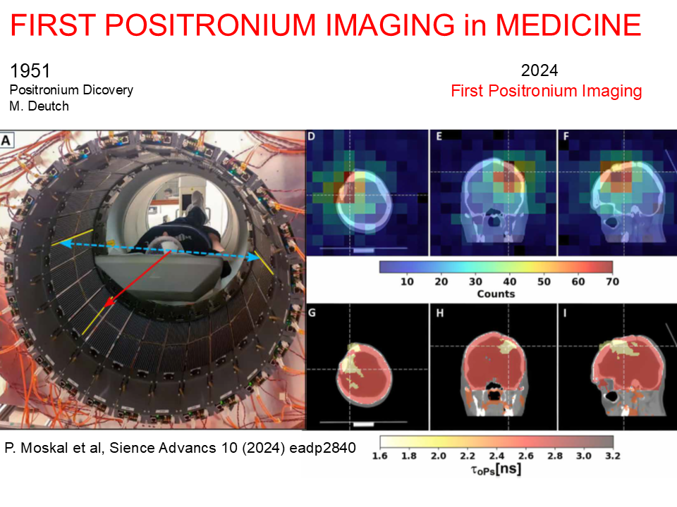
Fig. 2. Positronium imaging of a human brain.
(A) Photograph of the patient in the modular J-PET tomograph. The patient diagnosed with brain glioma was intravenously and intratumorally administered with pharmaceuticals labeled with the 68 Ga radionuclide, which emits positrons and prompt gamma rays (the course of imaging is illustrated in Fig. 1 above). The superimposed arrows represent photons from electron-positron annihilation (blue dashed arrow) and prompt gamma from the deexcitation of the 68 Zn* radionuclide (red solid arrow). The plastic strips of the tomograph in which the gamma rays interacted have been highlighted in yellow.
The figures and caption are taken from the article published in Science Advances, where more details are available.
https://www.science.org/doi/10.1126/sciadv.adp2840